


 Foundational characteristics of cancer include proliferation, angiogenesis, migration, evasion of apoptosis, and cellular immortality. Find key markers for these cellular processes and antibodies to detect them.
 The SUMOplot™ Analysis Program predicts and scores sumoylation sites in your protein. SUMOylation is a post-translational modification involved in various cellular processes, such as nuclear-cytosolic transport, transcriptional regulation, apoptosis, protein stability, response to stress, and progression through the cell cycle.
 The Autophagy Receptor Motif Plotter predicts and scores autophagy receptor binding sites in your protein. Identifying proteins connected to this pathway is critical to understanding the role of autophagy in physiological as well as pathological processes such as development, differentiation, neurodegenerative diseases, stress, infection, and cancer.
Anti-MMP2 / Collagenase Type IV A Antibody
Mouse Monoclonal Antibody
- SPECIFICATION
- CITATIONS
- PROTOCOLS
- BACKGROUND

Application
| WB, IF, FC |
|---|---|
| Primary Accession | P08253 |
| Other Accession | 513617 |
| Reactivity | Human |
| Host | Mouse |
| Clonality | Monoclonal |
| Isotype | Mouse / IgG1 |
| Clone Names | MMP2/1501 |
| Calculated MW | 73882 Da |
| Gene ID | 4313 |
|---|---|
| Other Names | 72kD type IV collagenase; CLG4A; Collagenase Type 4 alpha; Collagenase type IV A; Gelatinase A; Gelatinase alpha; Gelatinase neutrophil; Matrix metalloproteinase-2; MMP2; MONA; Neutrophil gelatinase; PEX; TBE-1 |
| Application Note | Flow Cytometry (0.5-1ug/million cells); ,Immunofluorescence (0.5-1ug/ml); ,Western Blotting (0.5-1ug/ml) ,Optimal dilution for a specific application should be determined. |
| Format | 200ug/ml of Ab purified from Bioreactor Concentrate by Protein A/G. Prepared in 10mM PBS with 0.05% BSA & 0.05% azide. Also available WITHOUT BSA & azide at 1.0mg/ml. |
| Storage | Store at 2 to 8°C.Antibody is stable for 24 months. |
| Precautions | Anti-MMP2 / Collagenase Type IV A Antibody is for research use only and not for use in diagnostic or therapeutic procedures. |
| Name | MMP2 |
|---|---|
| Synonyms | CLG4A |
| Function | Ubiquitinous metalloproteinase that is involved in diverse functions such as remodeling of the vasculature, angiogenesis, tissue repair, tumor invasion, inflammation, and atherosclerotic plaque rupture. As well as degrading extracellular matrix proteins, can also act on several nonmatrix proteins such as big endothelial 1 and beta- type CGRP promoting vasoconstriction. Also cleaves KISS at a Gly-|-Leu bond. Appears to have a role in myocardial cell death pathways. Contributes to myocardial oxidative stress by regulating the activity of GSK3beta. Cleaves GSK3beta in vitro. Involved in the formation of the fibrovascular tissues in association with MMP14. [Isoform 2]: Mediates the proteolysis of CHUK/IKKA and initiates a primary innate immune response by inducing mitochondrial- nuclear stress signaling with activation of the pro-inflammatory NF- kappaB, NFAT and IRF transcriptional pathways. |
| Cellular Location | [Isoform 1]: Secreted, extracellular space, extracellular matrix. Membrane. Nucleus Note=Colocalizes with integrin alphaV/beta3 at the membrane surface in angiogenic blood vessels and melanomas. Found in mitochondria, along microfibrils, and in nuclei of cardiomyocytes |
| Tissue Location | Produced by normal skin fibroblasts. PEX is expressed in a number of tumors including gliomas, breast and prostate |

Thousands of laboratories across the world have published research that depended on the performance of antibodies from Abcepta to advance their research. Check out links to articles that cite our products in major peer-reviewed journals, organized by research category.
info@abcepta.com, and receive a free "I Love Antibodies" mug.

Provided below are standard protocols that you may find useful for product applications.
Background
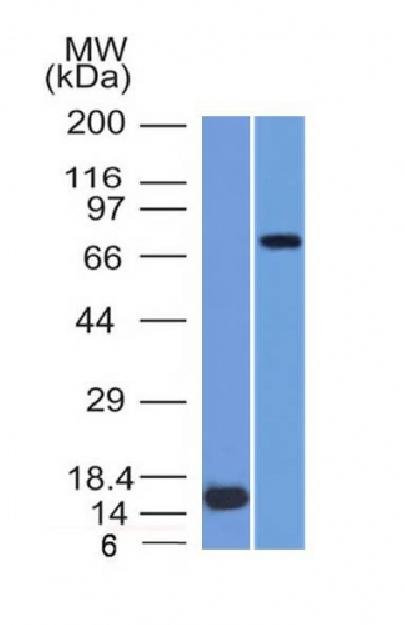
It recognizes a protein of 72kDa, which is identified as MMP2. The matrix metalloproteinases (MMP) are a family of peptidase enzymes responsible for the degradation of extracellular matrix components, including collagen, gelatin, Fibronectin, Laminin and proteoglycan. Transcription of MMP genes is differentially activated by phorbol ester, lipopolysaccharide (LPS) or staphylococcal enterotoxin B (SEB). MMP catalysis requires both calcium and zinc. MMP-2 (also designated type IV collagenase) cleaves collagen types IV,V, VII and X and gelatin type I. Activation of MMP-2 secretion requires the Ras signaling pathway.
If you have used an Abcepta product and would like to share how it has performed, please click on the "Submit Review" button and provide the requested information. Our staff will examine and post your review and contact you if needed.
If you have any additional inquiries please email technical services at tech@abcepta.com.
Ordering Information
Other Products
Shipping Information
